

If doctors want to know more about a patient’s risk of cardiovascular disease, they can order a cardiac stress test. But when it comes to stroke risk, there is no equivalent scalable and cost-effective test of brain function that can help doctors advise patients about their potential risk. A questionnaire asking patients about risk factors is currently the best tool for assessing such risk.
Now, a team of engineers and scientists at Caltech and USC’s Keck School of Medicine has developed a headset-based device that can non-invasively assess a patient’s stroke risk by monitoring changes in blood flow and volume while the participant holds their breath. The device features a laser-based system and has shown promising results in distinguishing between those at low and high risk of stroke.
New Technology Could Revolutionize the Way Stroke Risk is Assessed
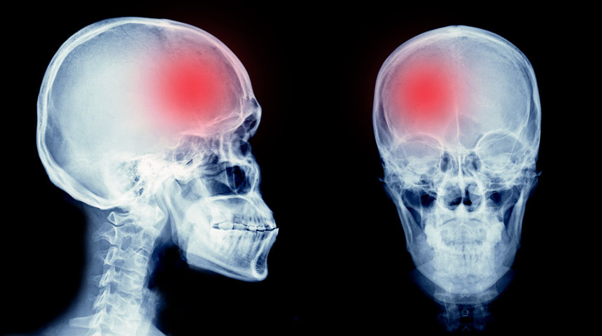
Around 1 in 1000 people suffer a stroke every year. The risk increases with age. On average, men are affected slightly more frequently. A stroke is caused by the blockage or rupture of an artery in the brain, which leads to a reduction in blood flow. The brain cells die quickly because they are no longer supplied with oxygen – about 2 million per minute during a stroke. Even if you survive the stroke, it can result in physical impairments such as speech and vision problems, impaired attention and concentration, paralysis on one side of the body or problems swallowing.

The researchers led by Simon Mahler, one of the lead authors of an article describing the new technique and device in the journal Biomedical Optics Express, and a postdoctoral fellow in the laboratory of Changhuei Yang, Thomas G. Myers Professor of Electrical Engineering, Bioengineering and Medical Engineering at Caltech and a researcher at the Heritage Medical Research Institute, have for the first time been able to use this device to determine whether or not someone is at significant risk of having a stroke in the future based on a physiological measurement. The experts believe this can truly revolutionize the way stroke risk is assessed and will ultimately help physicians determine whether a patient’s risk is stable or worsening.
“Our optical technology for noninvasive measurement of blood flow is expected to be useful for a number of applications in brain disease,” said Yang, who is also a senior staff scientist in electrical engineering at Caltech. He noted that this project is part of a larger collaboration with Dr. Charles Liu, professor of clinical neurological surgery, surgery, psychiatry and behavioral sciences, and biomedical engineering at USC’s Keck School of Medicine, and his team.
Optical Speckle Contrast Spectroscopy to Assess the Risk
In general, blood vessels become stiffer with age, meaning it is harder for them to dilate to allow blood to pass through. This in turn means that the person is more prone to strokes. The Caltech team has developed a compact device that shines infrared laser light through the skull and into the brain at one point and then uses a special camera nearby to collect the light that bounces back after being scattered by the blood flowing in the blood vessels. This approach, called speckle contrast optical spectroscopy (SCOS), measures the decrease in light intensity from the point where the light enters the skull to the point where the reflected light is collected to determine the volume of blood in the blood vessels of the brain. It also examines how the light is scattered and creates spots in the camera’s field of view. The spots fluctuate in the images depending on the blood flow rate in the blood vessels. The faster the blood flows, the faster the spot field changes.
Using these measurements, the researchers can calculate the relationship between the blood flow and the volume of blood flowing through the vessel to get an idea of the patient’s risk of stroke. The team conducted a study with 50 participants. They used the currently used stroke risk questionnaire, the Cleveland Stroke Risk Calculator, to divide the participants into two groups: one at low risk and one at high risk. Blood flow was then measured in each subject for three minutes to quantify the flow rate and volume of blood reaching the brain. After one minute, the participants were asked to hold their breath.
Holding the breath stresses the brain as it begins to realize that it is taking in too much carbon dioxide and too little oxygen. It goes into what Mahler calls “panic mode” and starts pumping oxygen from the rest of the body to itself. This greatly increases blood flow to the brain. As soon as you stop holding your breath, oxygen levels return to baseline. While this happens in both low and high stroke risk people, the researchers found that there were differences between the groups in terms of how the blood moved through the vessels. Using the SCOS technique, the researchers can measure how much the blood vessels expand while the subject holds their breath and how much faster the blood flows through the vessels in response. “According to the researchers, these reactive measurements are an indicator of vascular stiffness. This technology makes it possible for the first time to carry out this type of measurement non-invasively.
Technology With a Future
The researchers found clear and striking evidence of a different response in blood flow and blood volume between the two groups. In the low stroke risk group, they observed a smaller increase in blood flow during the breathing exercise compared to the high stroke risk group, but a larger increase in blood volume – an indication that more blood can flow through the dilated blood vessels. The researchers can clearly see that the higher risk group has a higher flow to volume ratio, meaning they have a faster flow but a lower blood volume during breath holding. This is caused by the stiffness of the blood vessels and indicates a higher risk of rupture. If someone comes to us with an extremely high flow-to-volume ratio, it is likely that they will suffer a stroke in the near future.
The team is conducting further studies with the current prototype imaging device on patients at a hospital in Visalia, California, to collect additional data from a larger, more diverse population. The researchers also plan to incorporate machine learning into the device’s data collection process and conduct a clinical trial following patients over a period of more than two years to improve the technology. They hope that the device can eventually be used not only for pre-stroke risk screening, but also to detect strokes that have already occurred.